Diuretics
General Pharmacology
 Renal
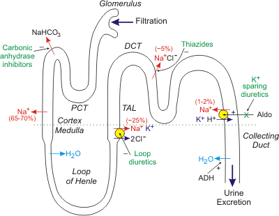
handling of sodium and water. To understand the action of diuretics, it
is first necessary to review how the kidney filters fluid and forms urine. As
blood flows through the kidney, it passes into glomerular capillaries located
within the cortex (outer zone of the kidney). These glomerular capillaries are
highly permeable to water and electrolytes. Glomerular capillary hydrostatic
pressure drives (filters) water and electrolytes into Bowman's space and into
the proximal convoluting tubule (PCT). About 20% of the plasma that enters the
glomerular capillaries is filtered (termed filtration fraction). The PCT, which
lies within the cortex , is the site of sodium, water and bicarbonate transport
from the filtrate (urine), across the tubule wall, and into the interstitium of
the cortex. About 65-70% of the filtered sodium is removed from the urine found
within the PCT (this is termed sodium reabsorption). This sodium is reabsorbed
isosmotically, meaning that every molecule of sodium that is reabsorbed is
accompanied by a molecule of water. Renal
handling of sodium and water. To understand the action of diuretics, it
is first necessary to review how the kidney filters fluid and forms urine. As
blood flows through the kidney, it passes into glomerular capillaries located
within the cortex (outer zone of the kidney). These glomerular capillaries are
highly permeable to water and electrolytes. Glomerular capillary hydrostatic
pressure drives (filters) water and electrolytes into Bowman's space and into
the proximal convoluting tubule (PCT). About 20% of the plasma that enters the
glomerular capillaries is filtered (termed filtration fraction). The PCT, which
lies within the cortex , is the site of sodium, water and bicarbonate transport
from the filtrate (urine), across the tubule wall, and into the interstitium of
the cortex. About 65-70% of the filtered sodium is removed from the urine found
within the PCT (this is termed sodium reabsorption). This sodium is reabsorbed
isosmotically, meaning that every molecule of sodium that is reabsorbed is
accompanied by a molecule of water.
As the tubule dives into the medulla, or
middle zone of the kidney, the tubule becomes narrower and forms a loop (Loop of
Henle) that reenters the cortex as the thick ascending limb (TAL) that travels
back to near the glomerulus. Because the interstitium of the medulla is very
hyperosmotic and the Loop of Henle is permeable to water, water is reabsorbed
from the Loop of Henle and into the medullary interstitium. This loss of water
concentrates the urine within the Loop of Henle. The TAL, which is impermeable
to water, has a cotransport system that reabsorbs sodium, potassium and chloride
at a ratio of 1:1:2. Approximately 25% of the sodium load of the original
filtrate is reabsorbed at the TAL. From the TAL, the urine flows into the distal
convoluting tubule (DCT), which is another site of sodium transport (~5% via a
sodium-chloride cotransporter) into the cortical interstitium (the DCT is also
impermeable to water). Finally, the tubule dives back into the medulla as the
collecting duct and then into the renal pelvis where it joins with other
collecting ducts to exit the kidney as the ureter. The distal segment of the DCT
and the upper collecting duct has a transporter that reabsorbs sodium (about
1-2% of filtered load) in exchange for potassium and hydrogen ion, which are
excreted into the urine. It is important to note two things about this
transporter. First, its activity is dependent on the tubular concentration of
sodium, so that when sodium is high, more sodium is reabsorbed and more
potassium and hydrogen ion are excreted. Second, this transporter is regulated
by aldosterone, which is a mineralocorticoid hormone secreted by the adrenal
cortex. Increased aldosterone stimulates the reabsorption of sodium, which also
increases the loss of potassium and hydrogen ion to the urine. Finally, water is
reabsorbed in the collected duct through special pores that are regulated by
antidiuretic
hormone, which is released by the posterior pituitary. ADH increases the
permeability of the collecting duct to water, which leads to increased water
reabsorption, a more concentrated urine and reduced urine outflow (antidiuresis).
Nearly all of the sodium originally filtered is reabsorbed by the kidney, so
that less than 1% of originally filtered sodium remains in the final urine.
Mechanisms of diuretic drugs. Diuretic drugs increase urine
output by the kidney (i.e., promote diuresis). This is accomplished by altering
how the kidney handles sodium. If the kidney excretes more sodium, then water
excretion will also increase. Most diuretics produce diuresis by inhibiting the
reabsorption of sodium at different segments of the renal tubular system.
Sometimes a combination of two diuretics is given because this can be
significantly more effective than either compound alone (synergistic effect).
The reason for this is that one nephron segment can compensate for altered
sodium reabsorption at another nephron segment; therefore, blocking multiple
nephron sites significantly enhances efficacy.
Loop diuretics inhibit the sodium-potassium-chloride cotransporter
in the thick ascending limb (see above figure). This transporter normally
reabsorbs about 25% of the sodium load; therefore, inhibition of this pump
can lead to a significant increase in the distal tubular concentration of
sodium, reduced hypertonicity of the surrounding interstitium, and less
water reabsorption in the collecting duct. This altered handling of sodium
and water leads to both diuresis (increased water loss) and natriuresis
(increased sodium loss). By acting on the thick ascending limb, which
handles a significant fraction of sodium reabsorption, loop diuretics are
very powerful diuretics. These drugs also induce renal synthesis of
prostaglandins, which contributes to their renal action including the
increase in renal blood flow and redistribution of renal cortical blood
flow.
Thiazide diuretics, which are the most commonly used diuretic,
inhibit the sodium-chloride transporter in the distal tubule. Because this
transporter normally only reabsorbs about 5% of filtered sodium, these
diuretics are less efficacious than loop diuretics in producing diuresis and
natriuresis. Nevertheless, they are sufficiently powerful to satisfy most
therapeutic needs requiring a diuretic. Their mechanism depends on renal
prostaglandin production.
Because loop and thiazide diuretics increase sodium delivery to the
distal segment of the distal tubule, this increases potassium loss
(potentially causing hypokalemia) because the increase
in distal tubular sodium concentration stimulates the aldosterone-sensitive
sodium pump to increase sodium reabsorption in exchange for potassium and
hydrogen ion, which are lost to the urine. The increased hydrogen ion loss
can lead to metabolic alkalosis. Part of the loss of potassium
and hydrogen ion by loop and thiazide diuretics results from activation of
the
renin-angiotensin-aldosterone system that occurs because of reduced
blood volume and arterial pressure. Increased aldosterone stimulates sodium
reabsorption and increases potassium and hydrogen ion excretion into the
urine.
There is a third class of diuretic that is referred to as
potassium-sparing diuretics. Unlike loop and thiazide diuretics, some of
these drugs do not act directly on sodium transport. Some drugs in this
class antagonize the actions of aldosterone (aldosterone receptor
antagonists) at the distal segment of the distal tubule. This causes
more sodium (and water) to pass into the collecting duct and be excreted in
the urine. They are called K+-sparing diuretics because they do
not produce hypokalemia like the loop and thiazide diuretics. The reason for
this is that by inhibiting aldosterone-sensitive sodium reabsorption, less
potassium and hydrogen ion are exchanged for sodium by this transporter and
therefore less potassium and hydrogen are lost to the urine. Other
potassium-sparing diuretics directly inhibit sodium channels associated with
the aldosterone-sensitive sodium pump, and therefore have similar effects on
potassium and hydrogen ion as the aldosterone antagonists. Their mechanism
depends on renal prostaglandin production. Because this class of diuretic
has relatively weak effects on overall sodium balance, they are often used
in conjunction with thiazide or loop diuretics to help prevent hypokalemia.
Carbonic anhydrase inhibitors inhibit the transport of bicarbonate
out of the proximal convoluted tubule into the interstitium, which leads to
less sodium reabsorption at this site and therefore greater sodium,
bicarbonate and water loss in the urine. These are the weakest of the
diuretics and seldom used in cardiovascular disease. Their main use is in
the treatment of glaucoma.
Cardiovascular effects of diuretics. Through their effects on
sodium and water balance, diuretics decrease blood volume and venous pressure.
This decreases cardiac filling (preload)
and, by the
Frank-Starling mechanism, decreases ventricular
stroke volume
and cardiac
output, which leads to a fall in arterial pressure. The decrease in venous
reduces capillary
hydrostatic pressure, which decreases
capillary fluid
filtration and promotes capillary fluid reabsorption, thereby reducing
edema if
present. There is some evidence that loop diuretics cause venodilation, which
can contribute to the lowering of venous pressure. Long-term use of diuretics
result in a fall in
systemic vascular resistance (by unknown mechanisms) that helps to sustain
the reduction in arterial pressure.
|

